
Fibroid Treatment · Uterus-Preserving Surgery · Şişli, Istanbul
Fibroid Surgery in Istanbul: Uterus-Preserving Surgery
The number, size and position of fibroids matter, but so do your symptoms and pregnancy plans. Together with Prof. Dr. Hanifi Şahin, review monitoring, medication and open, laparoscopic, hysteroscopic or vaginal surgical options.
- Surgical experience of removing more than 200 fibroids from a single uterus
- Aiming to preserve the uterus even in complex cases, including a giant 60 cm, 16.8 kg fibroid
- Not every fibroid needs surgery: monitoring, medication and surgical options are discussed together
- Send your reports and we can do an initial assessment by phone or WhatsApp
Appointment line 24/7 · You can send your reports via WhatsApp.
- Prof. Dr. Hanifi ŞahinGynecologic oncology surgery subspecialist
- 200+ fibroidsRemoved from a single uterus
- IHA · TGRT HaberCases reported in the press
- 24/7 appointment linePhone and WhatsApp
The first questions about fibroids
Are fibroids always dangerous?
No. Fibroids are mostly benign masses and some never cause symptoms; they only need regular follow-up.
Read more →Does every fibroid need surgery?
No. Treatment is chosen according to symptoms, bleeding and anaemia, growth rate, the location of the fibroid, age and pregnancy plans.
Read more →Is treatment possible without removing the uterus?
In many patients a uterus-preserving myomectomy can be performed. Suitability depends on the anatomy of the fibroids and surgical safety.
Read more →Which method is the best?
There is no single “best” method for everyone. Monitoring, medication, hysteroscopy, laparoscopy, open or vaginal surgery are chosen for the individual patient.
Read more →Three questions that decide the plan
The sections below explain how the decision is made, from the first examination to the surgical route.
Do I need treatment at all?
Monitoring or treatment depends on symptoms and the effect of the fibroid on your life.
Read more →Which surgical route?
Hysteroscopic, laparoscopic, vaginal or open surgery: how the route is chosen.
Read more →Can my uterus be preserved?
What “uterus-preserving” means, and what it does not guarantee.
Read more →An organ-preserving approach and advanced pelvic surgery experience in fibroid surgery
Prof. Dr. Hanifi Şahin is a specialist in obstetrics and gynecology who trained in gynecologic oncology surgery. His knowledge of advanced pelvic anatomy allows detailed planning aimed at preserving the uterus even with numerous or large fibroids.
Gynecologic oncology training
Advanced pelvic surgery training at Başkent University and Ankara Dr. Zekai Tahir Burak Training and Research Hospital.
Aim of preserving the uterus
The map of fibroids, pregnancy plans and surgical safety are considered together.
The method fits the patient
Hysteroscopic, laparoscopic, vaginal or open surgery; no single method suits everyone.
Transparent information
Advantages, risks and alternatives are explained verbally and in writing before surgery.
Surgical experience with fibroids of different sizes and numbers
Every fibroid case is different. The cases below are shared to show how the surgical plan can change according to the number, size and position of the fibroids.
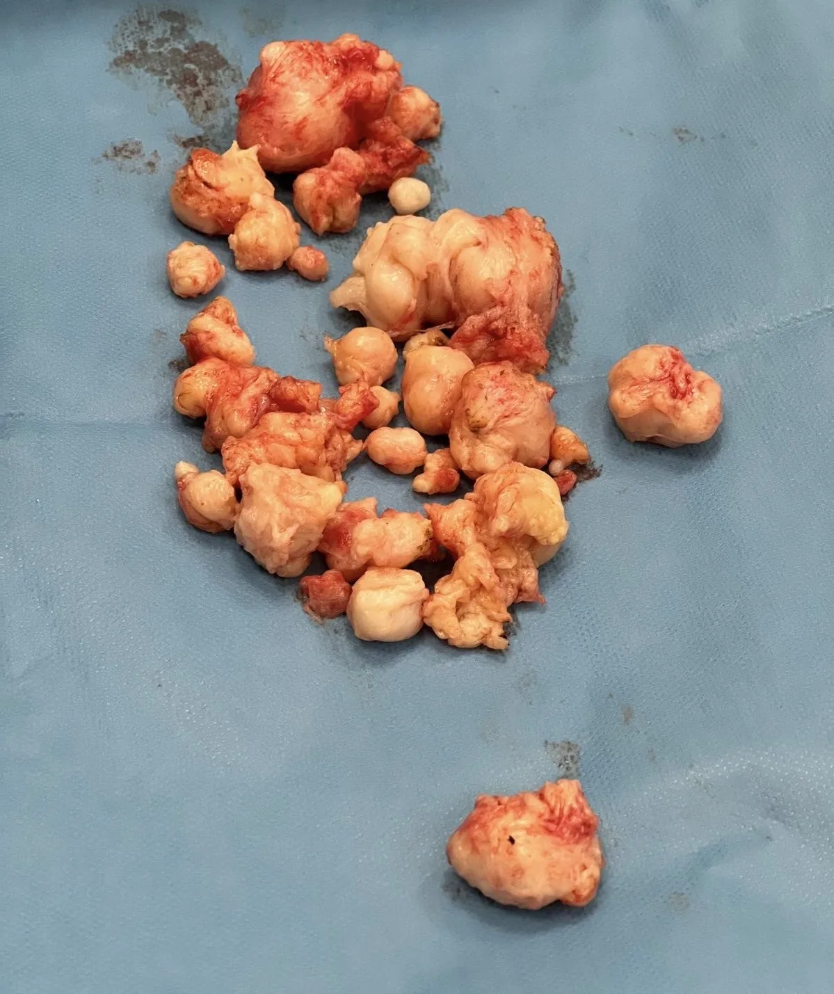
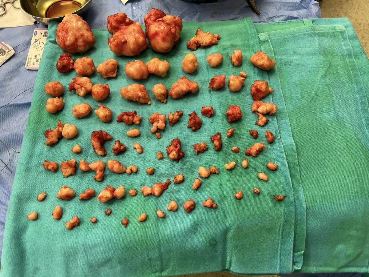
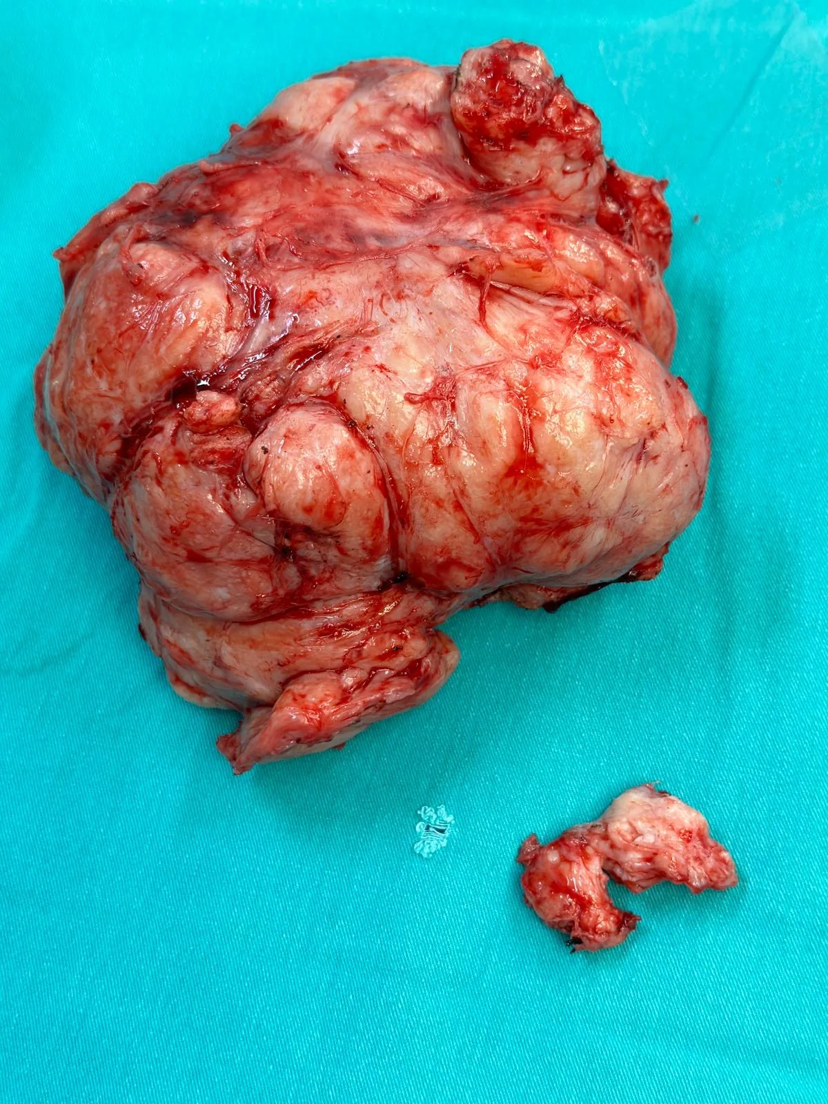
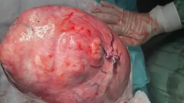
These examples belong to specific patients; results vary from person to person and do not imply that the same method or result will be suitable for another patient.
A clear path from enquiry to treatment
- Initial informationConsultation details are shared by phone or WhatsApp; you can send the reports you have.
- Examination and imagingYour symptoms, ultrasound/MRI findings and pregnancy plans are assessed together.
- Personal treatment planMonitoring, medication or surgical options are planned and explained with their advantages and risks.
- Surgery and follow-upRecovery after surgery, the pathology result and the waiting time before pregnancy are discussed.
Please bring with you
- Digital copies of your ultrasound and MRI images
- Imaging reports
- Recent blood count and iron results
- A list of the medication and supplements you use
- If you have had previous surgery, the operation note and pathology result
What are uterine fibroids?
A fibroid (leiomyoma, myoma) is a mostly benign mass that develops from the smooth muscle cells and connective tissue that make up most of the uterus. There may be a single fibroid or many of different sizes. Some fibroids stay the same size for years, while others grow with hormones, pregnancy or age.
A diagnosis of fibroids does not by itself mean suspected cancer or the need for surgery. What determines the need for treatment is the effect the fibroid has on the person: heavy bleeding, anaemia, pain or pressure, urinary or bowel symptoms, or an effect on pregnancy plans.
Types of fibroids and why location matters
Fibroids are classified by where they lie in the uterus, and one patient may have several types at once. Location influences bleeding, pain, pressure, pregnancy and the surgical route that can be used.
- Submucosal fibroids grow towards the uterine cavity. Even when small they can cause heavy or intermenstrual bleeding or fertility problems. Suitable ones can be removed hysteroscopically, through the cervix.
- Intramural fibroids lie within the muscle wall. Depending on their size and closeness to the cavity they can cause bleeding, pain or pressure.
- Subserosal fibroids grow towards the outside of the uterus. They tend to cause pressure symptoms rather than bleeding.
- Pedunculated and cervical fibroids are rarer forms that may need a different technical approach.
What are the symptoms of fibroids?
A large proportion of fibroids cause no symptoms. When they do, complaints depend on the location, number and size of the fibroids:
- Periods that are heavier or longer than before; clots, bleeding between periods or frequent periods
- Tiredness, palpitations, breathlessness or dizziness caused by iron deficiency and anaemia
- Pain or pressure in the groin, lower back or lower abdomen
- Abdominal enlargement, fullness or a palpable firmness
- Frequent urination or incomplete bladder emptying because of pressure on the bladder; constipation
Any new vaginal bleeding after menopause should always be assessed.
How are fibroids diagnosed?
The assessment starts with your complaints, menstrual pattern, pregnancy plans and previous treatments. Imaging then shows the number, size and position of the fibroids.
- Ultrasound is the first-line method in most patients. Vaginal ultrasound shows the cavity and muscle wall in detail; abdominal ultrasound can be added for large masses.
- MRI is used when the map of the fibroids must be clarified, when there are many or very large fibroids, or when the picture is unusual.
- Hysteroscopy examines the uterine cavity directly when a submucosal fibroid or polyp is suspected.
A useful report does not only say “fibroid present”; it gives the number, three dimensions, position and relation to the cavity of each fibroid.
When to monitor and when to treat
Finding a fibroid does not mean that treatment must start immediately. Fibroids that cause no symptoms, have typical imaging features and do not grow significantly can be followed at regular intervals. The frequency of follow-up depends on age, size and position, earlier scans, menopausal status and complaints.
The main reasons to consider treatment are heavy bleeding that impairs quality of life, anaemia despite treatment, marked pain or pressure, urinary or bowel complaints, fibroids that affect the uterine cavity, and fibroids that interfere with pregnancy plans. A rapidly growing fibroid or an unusual appearance on imaging needs a separate assessment.
Treatment options
The aim of treatment is not always to remove the fibroid completely. In some patients the goal is to control bleeding and correct anaemia, in others to relieve pressure, correct the uterine cavity or find a path that suits pregnancy plans. Clarifying the goal helps avoid unnecessary treatment and choose the right method.
| Approach | Main aim | Uterus kept? |
|---|---|---|
| Regular monitoring | Following a fibroid with no or mild symptoms | Yes |
| Medication for symptoms | Reducing bleeding and pain, correcting anaemia | Yes |
| Hysteroscopic surgery | Removing fibroids that grow into the cavity | Yes |
| Myomectomy (laparoscopic, vaginal or open) | Removing the fibroids and repairing the uterus | Yes |
| Non-surgical procedures (e.g. embolisation, HIFU) | Reducing fibroids without surgery in selected patients | Yes (with limits) |
| Hysterectomy | Definitive treatment when pregnancy is not planned | No |
Fibroid surgery and how the route is chosen
When people say “fibroid surgery” they usually mean myomectomy: removing the fibroids and repairing the uterus. Myomectomy is the basic surgical approach for suitable patients who want to keep the uterus or plan a pregnancy. The aim is not only to take out the visible mass, but also to control bleeding, repair the muscle wall safely, protect the cavity and respect neighbouring organs.
The route is not chosen on the patient’s wish alone. The map of the fibroids, their distance from the cavity and the outer surface, relation to vessels, the patient’s earlier surgery and pregnancy plans all matter.
- Hysteroscopic: for fibroids growing into the cavity; no incision in the abdomen.
- Laparoscopic (keyhole): small entry points in the abdomen, removal of the fibroids and repair of the uterus. Suitable for many, but not every, fibroid.
- Vaginal (V-NOTES): in selected cases, surgery through the vagina without abdominal scars.
- Open: may be the safest option for very large or very numerous fibroids, or when other routes are unsuitable.
There is no universally “better” route. A closed operation is not automatically superior to an open one if it makes the repair of the uterus less safe.
Uterus-preserving fibroid surgery
Uterus-preserving surgery aims, when it is possible and safe, to remove the fibroids and rebuild the anatomical integrity of the uterus. It is not only for patients who want a child soon: a patient who wishes to keep her uterus without a pregnancy plan should have this preference included in the decision, within medical safety limits.
“Preserving the uterus” is not the same as “guaranteeing fertility”. Myomectomy leaves the uterus in place, but age, ovarian reserve, tubes, partner factors, intrauterine adhesions and other conditions also determine the chance of pregnancy. The waiting time before pregnancy and the mode of delivery after surgery are decided individually.
Prof. Dr. Hanifi Şahin’s training in gynecologic oncology surgery, with advanced pelvic anatomy and bleeding control, supports the planning of such operations in complex patients.
Fibroids, fertility and pregnancy
Many women with fibroids conceive naturally and have healthy pregnancies. The effect on fertility is not the same in every patient. Submucosal fibroids that distort the cavity, masses near the openings of the tubes, and some large intramural fibroids matter more, while subserosal fibroids usually have a more limited effect. If a woman has difficulty conceiving, a fibroid does not mean it is certainly the cause: age, ovulation, tubes and partner factors are also assessed.
Preparation and recovery
When surgery is decided, preparation aims to reduce risk and clarify the goal. Current ultrasound or MRI is reviewed, the map of fibroids is drawn, and blood count and iron stores are checked. If there is marked anaemia, iron treatment, medication to control bleeding or, in selected patients, temporary hormonal suppression can be planned. Tell the team about prescription medication, blood thinners, herbal products, allergies and previous operations.
Recovery depends on the method, the number of fibroids removed, the depth of the repair, blood loss and general health. Return to daily life is usually shorter after hysteroscopy and earlier after laparoscopy than after open surgery, but small external incisions do not mean the repair inside the uterus is small. Tiredness, abdominal bloating and light vaginal bleeding are common early on. Follow-up visits review the wound, the pathology result, bleeding pattern, anaemia, return to activity, the personal waiting time before pregnancy and long-term ultrasound follow-up.
Fibroids and menopause
With menopause, the fall in oestrogen and progesterone can make many fibroids shrink and bleeding stop, so for some women close to menopause with mild complaints monitoring is reasonable. However, it is not true that all fibroids disappear at menopause: pressure symptoms may persist, and any new vaginal bleeding after menopause must always be assessed.
How a consultation for fibroids works
The aim of the consultation is not to recommend surgery straight away, but to define the source of the complaints and the patient’s goals. At Prof. Dr. Hanifi Şahin’s clinic in Istanbul, existing ultrasound and MRI images, the history of periods and bleeding, previous treatments, pregnancy plans and any earlier operation notes are assessed together. At the first meeting the location of the fibroids and their relation to the cavity are explained, and the monitoring, medication, non-surgical and surgical options that are suitable for assessment are discussed. If surgery is considered, hysteroscopic, laparoscopic, vaginal or open routes are compared with their advantages and risks. A second opinion on an operation recommended elsewhere is also possible.
Sources and content principles
This guide is written to help patients understand the basic concepts and treatment options for fibroids. It does not replace a personal examination, diagnosis or treatment advice; medical decisions must be made with current clinical findings and the patient’s preferences.
- American College of Obstetricians and Gynecologists (ACOG): Uterine Fibroids
- NICE Guideline NG88: Heavy menstrual bleeding – assessment and management
- Royal College of Obstetricians and Gynaecologists (RCOG): Uterine Artery Embolisation in the Management of Fibroids
What patients ask
At what size does a fibroid need surgery?
There is no single size limit. The decision depends on symptoms (bleeding, anaemia, pain, pressure), growth, location and pregnancy plans; a small submucosal fibroid can matter more than a larger subserosal one.
Can a fibroid of 10 cm or more be removed by keyhole surgery?
Sometimes. It depends on the number and position of the fibroids, the anatomy and surgical safety; in some large or numerous fibroids an open operation may be the safer route. The decision is made after examination and imaging.
Does a fibroid shrink by itself?
Some fibroids shrink, especially after menopause; others stay the same or grow. Follow-up and symptoms guide the decision.
Can a fibroid turn into cancer?
Fibroids are almost always benign. Cancers of the uterine muscle are rare, and a fibroid does not usually “turn into” cancer. A fibroid that grows quickly or looks unusual on imaging needs an additional assessment.
Do fibroids make it harder to get pregnant?
Not always. Fibroids that distort the cavity or lie near the tubes are more likely to matter. Other causes of difficulty conceiving are assessed too.
Is the uterus removed during fibroid surgery?
Not necessarily. Where it is safe and possible, myomectomy removes the fibroids and repairs the uterus. A hysterectomy is considered when pregnancy is not planned and the patient prefers definitive treatment, or when conservative surgery is unsafe.
Is keyhole or open surgery better?
Neither is better for everyone. The route is chosen by the fibroids’ number, size and position, safety of repair and the patient’s history.
How long is the hospital stay and when can I return to work?
It varies with the method; after hysteroscopy often the same day, after laparoscopy usually a short stay, and after open surgery a longer stay and recovery. Your surgeon will give you an individual plan.
Is there a herbal treatment for fibroids?
There is no reliable evidence that herbal products shrink fibroids. Some products can interact with medication; tell your doctor about anything you take.
Can I apply from abroad?
Yes. You can send your imaging and reports for an initial assessment; a physician’s examination is needed before any decision on surgery.
Let us review your fibroid reports together
Bring your ultrasound or MRI images and reports to the consultation, or send them on WhatsApp; let us discuss the options with their advantages and risks.
- Appointment line (24/7)+90 (538) 469 81 61
- WhatsAppSend a message
- ClinicCumhuriyet Mah., Halaskargazi Cad., Selamet Apt. No: 93 Kat: 2, Şişli / İstanbul
Open in maps